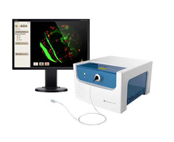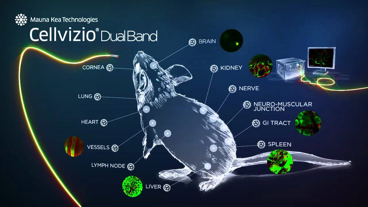
The research infrastructure if our Institute has just been expanded with high-end equipment: Cellvizio® Dual Band System by Manua Kea Technologies for in vivo imaging in pre-clinical trials at cell resolution.
Cellvizio® System is a fluorescence-based confocal laser endomicroscopy (CLE) system, one of two devices of its type currently in use in Poland. The system allows for real-time imaging of tissues and cells by means on non- or minimally invasive methods.
The System combines confocal microscope and a flexible confocal microprobe to take advantage of both technological solutions. Highly-sensitive detectors enable surface imaging in contact with the tissue as well as internal imaging by inserting the detector into the soft tissue of interest. Confocal imaging, in turn, allows for imaging at cell resolution which is unavailable in other in vivo imaging systems (MRI, CT, PET, SPECT, Ultrasound, among others).
What is particularly valuable about real-time in vivo endomicroscopy is:
• the access to any place of interest to the experimenter during the whole imaging process
• opportunity to capture functional and morphological images both in animals that are awake after previous immobilization, and in the animal under inhalation anaesthesia (light anaesthesia)
• possibility of dynamic changes recording in vivo in the same animal over a longer period
Such features of the equipment allow for a wide range of research, including:
• vascularisation assessment: its density, morphology and functioning,
• dynamic recording of cellular and molecular interactions
• analysis of biodistribution of medication, endocrine and paracrine substances,
• monitoring of physiological and functional changes in the anatomical context

MMRI PAS Cellvizio® System is currently located at the Department of Renal and Body Fluid Physiology,
supervised by Prof. Elżbieta Kompanowska-Jezierska MD PhD:
tel. 22 60 86 546; E-mail: This email address is being protected from spambots. You need JavaScript enabled to view it.
All the interested external and internal researchers are invited to contact the equipment supervisor.
Moreover, we are currently searching for the equipment operator: a person willing to undergo the training provided by the producer, who will next be responsible for the equipment exploitation, the design of protocols of staining of particular organs and tissues, research material preparation, performance of the imaging, deconvolution and confocal image analysis.
Interested persons are asked to contact the equipment supervisor.


